Skin Color and Tanning
Melanocytes are specialized cells in which biosynthesis of melanin takes place. During embryonic development in humans, presursor melanocytes arise from the neural crest cells and actively migrate to peripheral sites, especially the dermoepidermal junction. Melanin pigment is synthesized in specialized cytoplasmic organelles called 'melanosomes'. Within the melanosome, the enzymatic conversion of the amino acid tyrosine to melanin is catalyztd by the enzyme tyrosinase. All melanocytes have the ability to form malignant melanomas. Click here for more information on skin cancers.

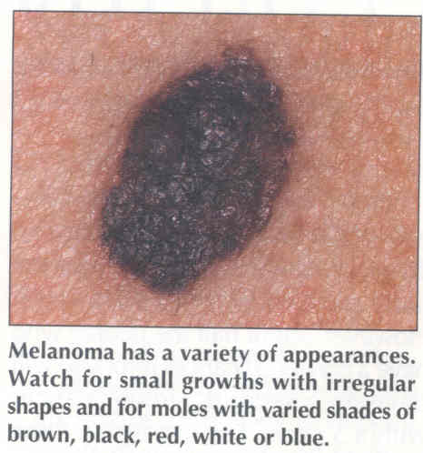
The photographs to the right and left are both melanomas. Notice the characteristic features: irregular border or edge; brown to black in color possibly with areas of white at the periphery; not raised but flush with the surface of the skin. Do not ignore any types of pigmented spots that have these characteristics.
The number of melanocytes in the skin varies depending upon the area of the body. Large numbers are found in the genitalia, face, oral and nasal mucosa whereas there are relatively few in the abdominal skin. There is very little variation in the number of melanocytes between people of different races. Thus, racial pigmentation differences do not depend upon the number of melanocytes but on the intensity of pigment produced by these cells.
Color variation in human skin derives chiefly from the presence within the epidermis of specialized melanin-bearing organelles, the melanosomes. Tanning of human skin on exposure to ultraviolet radiation (UVR) results from the presence of increased amounts of melanin within the epidermis. Melanosomes synthesized by melanocytes are passed to keratinocytes and transported within them to the epidermal surface.
Melanin pigmentation of human skin is divisible into two components:(1)constitutive skin color which is genetically programmed and is the amount of pigment the skin contains without exposure to UVR; (2)facultative (inducible) skin color which is the increased pigmentation seen as tanning following exposure to UVR. Skin hyperpigmentation can also result from hormonal changes, as seen during pregnancy. Therefore, a person's skin pigmentation arises from the complex interplay of their genetic constitution, hormones and exposure to UVR. In mice, more than 147 genes affect skin and hair color. Genetic influences on human melanin pigmentation may be equally complex.
Two types of melanin can be synthesized, the yellow-red 'pheomelanins' and the brown-black 'eumelanins'. Melanin is synthesized by melanocytes in membrane-bound sacs called melanosomes which are then transferred to keratinocytes. The synthesis of melanin, involving the conversion of the amino acid tyrosine into either black-brown eumelanin or red-yellow pheomelanin, or both, occurs in the melanosome of the melanocyte. The initial steps are the conversion of tyrosine into dopa (dihydroxyphenylalanine) and then dopa's conversion into dopaquinone, with both steps being catalyzed by tyrosinase. Dopaquinone then enters the pathway for pheomelanin or eumelanin synthesis.
The epidermal melanin is synthesized in the melanocyte and passed to the keratinocyte which migrates to the skin surface and degenerates to form the stratum corneum. The primary function of melanin is photoprotection. In the skin, melanin protects against the adverse effects of ultraviolet radiation by dissipating light energy as heat or in a chemical reaction. These photo-reactions of melanin can produce unstable free radicals which can then damage the cell. This is a particular problem with pheomelanin and may be relaed to the increased risk of cancer in people with increased levels of the red-yellow type of melanin.
The increase in melanin pigmentation after exposure of human skin to sunlight or to UVR from artificial sources is commonly known as 'tanning'. Tanning involves two distince biologic phenomena: (1)immediate tanning and (2)delayed tanning. Innediate tanning is optimally produced by both long UVR (320 to 380 nm) and visible (400 to 700 nm) light, and delayed tanning is optinally stimulated by exposure to the so-called sunburn spectrum (290 to 320 nm) and to a lesser extent by exposure to long-wave UVR and visable radiation.
Immediate tanning can best be seen in moderately to heavily pigmented individuals or in the previously exposed (tanned) areas of fair-skinned people. The skin begins to become hyperpigmented within 5 to 10 min on exposure to the midday summer sun and is maximally pigmented after 1 hour of irradiation. The hyperpigmented areas, when withdrawn from exposure to light, fade rapidly within the first 30 min, and thereafter the color usually fades gradually, so that after 3 to 4 hours the irradiated areas are barely hyperpigmented. Sometimes, after prolonged sun exposure (90 to 120 min), residual hyperpigmentation may be visible for as long as 24 to 36 hours, after which time newly synthesized melanin (delayed tanning) begins to pigment the skin.
Delayed tanning involves new production of melanosomes and therefore appears slowly over a period of days after exposure to UVR. An increased tyrosinase reaction is seen at 48 to 72 hours following exposure. No increased tyrosinase reactions are found immediately after the initial exposure.
The immediate tanning reaction appears to be based on the passing of preformed melanosomes to keratinocytes. Immediate tanning provides no photoprotection.
Hyperpigmentation of the skin in delayed tanning is due to the following changes in the normal process of melanin pigmentation: (1)An increase in the number of melanocytes by division and recruitment of dormant melanocytes; (2)increased production of melanosomes; (3)increased transfer of melanosomes; (4)activation of tyrosinase.

On the left: In the middle: On the right:
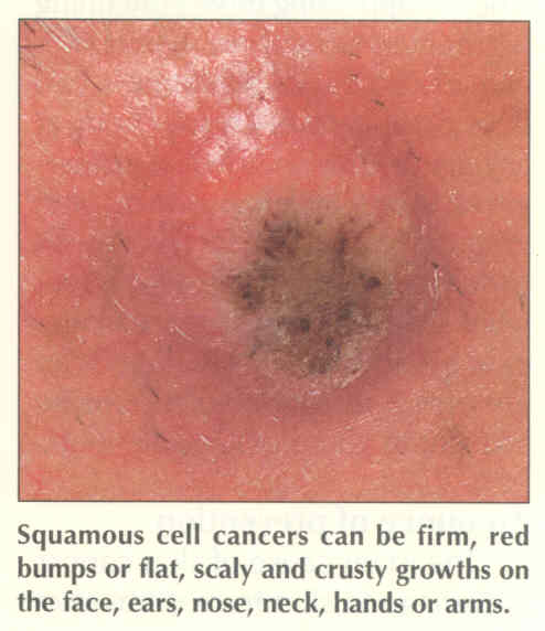
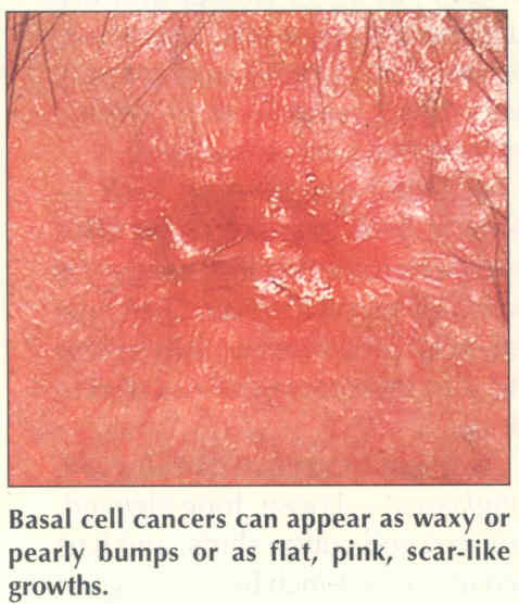
Basal Cell Squamous Cell Malignant
Carcinoma Carcinoma Melanoma
Always Wear Your Sun-Screen.